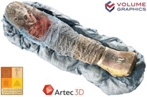
If you are in the field of analyzing ancient Egyptian mummies, here something really cool and informative. In the Sherit mummy project, researchers benefited from adding the results from a 3D scanner to a CT scan.
Sherit is a mummified Egyptian child who died 2,000 years ago. Even though the Rosicrucian Egyptian Museum in San Jose, California had the mummy since 1930, it only had a CT scan performed in 2005 to obtain more information about Sherit.
That analysis revealed the mummy to be a girl who was between four and a half and six years old when she died. Further information from the CT scan revealed that her body was wrapped in fine linen and covered with round earrings, an amulet, and a Roman period necklace, and allowed physicians to conclude that she probably died from dysentery or meningitis.
In spite of this rather extensive internal information, it did not include a full-color 3D image of the outer surface of the mummy.
With Volume Graphics Version 3.1 software and data from an Artec Eva 3D scanner and a Siemens Axiom CT scanner, the two datasets were merged with a click of a button. Unlike previous analysis, the result is a digital copy of the mummy, which shows how it looks on the outside as well as the inside.
Timing is an issue in this type of analysis since the museum that houses ancient treasures including mummies, usually puts a time limit on allowing uncontrolled exposure to the ambient and access by outsiders. The operator had 30 minutes to complete the scanning but it was done much faster – in less than 10 minutes allowing additional confirmation analysis to be performed.


Leave a Reply
You must be logged in to post a comment.